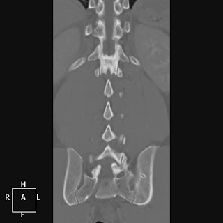
While we're on the subject of supernumerary rib oddities...
...unbelievably, there have been case reports in the literature of fully-formed ribs arising from the sacrum and even the coccyx, as in this case report from 1978 (below).
This particular example belongs to a 55Y woman who had spent her whole life unaware that she had a perfectly formed rib, with a well-formed capitellum, tubercle, and body, arising from her terminal coccygeal segment and terminating in her gluteal region.
Of the few reports of sacral or coccygeal ribs, the majority are short projections, and some are even described as "digits." Below is an example of a small "digit" rib extending from the sacrum.
There has been no description of deleterious effects from sacrococcygeal ribs, and authors stress that they should not be further imaged or intervened upon in the absence of symptoms.
1. Pais M, Levine A, Pais S. "Coccygeal Ribs: Development and Appearance in Two Cases" AJR 131:164-166. July 1978
2. Rashid M, Khalid M, Malik N. "Sacral Rib: A Rare Congenital Abnormality" Acta Orthop. Belg. 74: 429-431 2008
September 30, 2011
September 29, 2011
The Lumbar Ribs (a.k.a. the gorilla bone)
Mammals tend to have a preserved total number of vertebrae per species, and humans are no exception. Occasionally, when there is a slight variation in the expression of the Hox genes that encode the differentiation of the lumbar vertebrae (and occipital bone), there results in an extra (13th) pair of ribs on a morphologically lumbar vertebra. This effectively results in 13 thoracic vertebrae and 4 lumbar vertebrae, but the total number of thoracic and lumbar vertebrae rarely changes.
Hox genes (anterior-posterior axis patterning) shows a special evolutionary conservation in the cervical spine, such that almost every mammal is constrained to seven cervical vertebrae, regardless of the length of the neck. There is speculation that this may be a result of simultaneous Hox effects in neural patterning in the upper cervical spine, and that cervical spine variation leads to nonviable neural variations. One study has found an associated increased risk of cancer in children with a cervical rib.
The lumbar ribs are a similar result of differential Hox expression. The presence of lumbar ribs in mice can even be used as an index of the teratogenicity of a substance... but the increased incidence of natural lumbar ribs seems to imply that the distal Hox variation has less serious associations with neural patterning than proximal variations.
As an incidental note, the gorilla (Gorilla gorilla gorilla), has a similar typical pattern of 17 total thoracic and lumbar vertebrae, but tends to have the distribution of 13 thoracic vertebrae and 4 lumbar vertebrae.
References:
1. Narita Y, Kuratan S."Evolution of the Vertebral Formulae in Mammals: A Perspective on Developmental Constraints" JOURNAL OF EXPERIMENTAL ZOOLOGY (MOL DEV EVOL) 304B:91–106 (2005)
2. Galis F. "Why Do Almost All Mammals Have Seven Cervical Vertebrae? Developmental Constraints, Hox Genes, and Cancer" JOURNAL OF EXPERIMENTAL ZOOLOGY (MOL DEV EVOL) 285:19–26 (1999)
3. Merks J, Smets A, et al."Prevalence of RIB anomalies in normal Caucasian children and childhood cancer patients" "European Journal of Medical Genetics Vol 48, Issue 2, April-June 2005, Pages 113-129.
September 27, 2011
The Petro-occipital Suture
The sutures in the cranial vault are well-known, but the sutures/fissures of the skull base are much less discussed, although they can be classic origins for certain lesions. One such example is the petro-occipital suture (highlighted below in red; the contralateral suture unmarked), which, not suprisingly, lies between the occipital bone and the petrous aspect of the temporal bone.
The suture links the jugular foramen posteriorly and the foramen lacerum anteriorly.
So why is this suture important? It is a classic location where chondrosarcomas of the skull base can arise (6% of skull-based lesions). The painless, locally-invasive tumor arises from remnants of embryonal chondrocytes within the petro-occipital fissure and typically expands upward into intracranial structures.
Radiologically, a destructive mass located at this fissure (heterogeneous enhancement on post contrast T1, hyperintense on T2; chondroid calcification on CT) is highly suspicious of this diagnosis. The T1 w/ contrast image below (from Reference 2) is an example of a chondrosarcoma arising from the left petro-occipital fissure (the arrowhead demonstrates the petro-occipital fissure on the right).
Incidentally, although the molecular mechanism of eventual petro-occipital suture ossification is similar to that of the cranial vault sutures, it is unclear why it remains relatively unossified in the nonpathological state until late in adulthood.
A case report of an amyloidoma involving the petro-occipital suture has been described as well (images below). [Reference 3].
It has also been suggested that Maffuci syndrome and Ollier syndrome may be associated with intracranial chondrosarcomas arising from the petro-occipital fissure (Ref. 4).
References:
1. Balboni AL, Estenson TL, et al. "Assessing Age-Related Ossification of the Petro-Occipital Fissure: Laying the Foundation for Understanding the Clinicopathologies of the Cranial Base." The Anatomical Record Part A 282A:38–48 (2005)
2. Connor SEJ, Leung R, Natas S. "Imaging of the petrous apex: a pictorial review." The British Journal of Radiology, 81 (2008), 427–435
3. Simoens WA, van den Hauwe L, et al."Amyloidoma of the Skull Base." AJNR Am J Neuroradiol 21:1559–1562, September 2000
4. Tibbs RE, Bowles AP, Raila FA. "Maffuci's Syndrome and Intracranial Chondrosarcoma." Skull Base Surgery Vol 7, no. 1. (1997)


