October 4, 2011

Paraspinal Musculature: The Perils of Cricket

Attaching inferiorly to the fibers of the iliolumbar ligament, superiorly to the lowest rib, and medially to the transverse processes of the L1-L4 vertebral bodies, the quadratus lumborum muscle is visible in virtually all MRI studies of the lumbar spine... at least the axial sequences.  The QL is also visible in virtually every abdominal/pelvis CT, too.


Since the anterior surface of the muscle forms the front of the thoracolumbar fascia, it remains relatively sequestered from abdominal processes that affect its anterior neighbors: the kidney and psoas muscle.


Most of the interest in the world today in the quadratus lumborum muscle comes from Australia, where there is concern that fast-bowl cricket is leading to injury of the L4 pars interarticularis injury (an injury also commonly seen in tennis players).  Radiologists there determined that an enlarged quadratus lumborum is associated with an ipsilateral L4 pars fracture, however, there is debate whether the asymmetry of the muscle is a source of stress on the vertebral body or may be just a defensive adapatation to stress on the vertebral body.

Incidentally, the QL is the muscle that allows to hold our body horizontally on one elbow (as below).


1. McGill S, Juker D, Kropf P. "Quantitative intramuscular myoelectric activity of quadratus lumborum during a wide variety of tasks" Clinical Biomechanics 11:3, 170-172 (April 2011).
2. Engstrom, C. M., Walker, D., Kippers, V. and Buckley, R.  "Quadratus lumborum asymmetry and pars interatricularis injury in cricket fast bowlers: A prospective MRI examination" (2000). Quadratus lumborum asymmetry and pars interatricularis injury in cricket fast bowlers: A prospective MRI examination. In: , 2000 Pre-Olympic Congress Book of Abstracts. International Congress on Sport Science, Sports Medicine and Physical Education, Brisbane, (191-192). 7-12 September 2000. 

October 3, 2011

Lumbosacral transitional vertebrae

The Castellvi system for organizing lumbosacral variations.
Useful only for classification, not prognostic significance.
The design of the spine is awkard enough as it is, with the majority of the body's weight being transferred through L5-S1, but as if to make an awkward system even a little more awkward, every so often congenital vertebral variations occur at the lumbosacral junction and confuses the typical layout (see Hox genes in the 9/29/2011 post).

There are two main possibilities for a lumbosacral transitional vertebrae:

- "lumbarization of S1"
- "sacralization of L5"

which seem to be two ways of looking at the same thing... except in the first situation you essentially end up with an extra lumbar vertebra ("L6"), and in the second, you end up with one too few lumbar vertebrae (L1-L4).  Note that, unlike the "gorilla bone" example from 9/29/2011, it's assumed that there are the normal 12 ribs in both these situations, but then again it's more common to have thoracolumbar variations if there is a lumbosacral variation...

... so the final result of all this madness is that the only absolutely sure way to know which variations you're dealing with in the spine is to count down from C2...

...and numbering of these segments is critical since they frequently get intervened upon, either by surgery or by anesthesia.


A recent study demonstrated that the iliolumbar ligament (described as "absent in 70%" in earlier studies), actually denotes the lowest lumbar vertebra, which is not always L5 and should not be used as a marker for L5. 

Some object to the use of "L6" as the best term for this lowest lumbarized S1 vertebra since there is no L6 nerve root.  If "L6" is used, then the S1 nerve root would pass out beneath it, and then the S2 nerve root would passes out below/through the S1 neuroforamina...... and the whole vertebra-nerve root thing goes cattywhompus.

It is claimed by some, however, that the L5 nerve root always passes out the "last mobile" segment of the spine, so in a patient with a lumbarized S1 like the one below, the last mobile level is L6-S2, and the functional L5 nerve root corresponds to the "L6" nerve root.  The point hold true for patients with a sacralized L5 as well, and the L4 nerve root serves the usual function of the L5 nerve root.  This point has been debated, and some feel that this nerve root also shares some aspects of S1... but as of today, no thorough study has determined which is correct (perhaps both).


One other side note: whether lumbosacral transitional vertebrae is related to low back pain is controversial. A recent study demonstrated that nearly a 1/3 of a normal population sample had at least minor changes of transitional lumbosacral vertebrae in the spine, implying that it may not be as closely associated with low back pain as was once thought. Another recent study positively correlated Castellvi types II and IV with an increase in low back pain.

---
1.  Apazidis A, Ricart P, et al. "The Prevalence of Transitional Vertebrae in the Lumbar Spine."  The Spine Journal 11 (2011) 858-862
2. Kim YH, Lee PB, et al. "Dermatome Variation of Lumbosacral Nerve Roots in Patients with Transitional Lumbosacral Vertebrae" Anesthesia & Analgesia 2008; 106:1279-83
3. Carrino JA, Campbell PD, et al.  "Effect of Spinal Segment Variants on Numbering Vertebral Levels at Lumbar MR Imaging"  Radiology, 259, 196-202 April 2011
4. Nardo L, Alizai H, Virayavanich W. "Lumbosacral Transitional Vertebrae: Association with Low Back Pain" November 2012 Radiology,265, 497-503.

October 2, 2011

The Pit and the Pyramid

The middle ear has more nooks and crannies than a gnome's cave (gnomes live in caves, right?), and these are places where all kind of nastiness can hide out.  One example of such a hiding place is the sinus tympani, tucked up in the superior, posterior, and medial wall of the middle ear behind the pyramidal eminence.  The importance of the sinus tympani for radiologists is because this little nook is not always well visualized by ENT endoscopy, and it's a little cranny where acquired cholestomas, specifically the pars tensa cholestetomas, like to originate.

Hi res axial CT of the IAC

Hi res coronal CT of the IAC

There is variation in the depth of the sinus tympani from nearly flat with virtually no sinus at all, to a deep, hidden pocket... which of course is the more dangerous end of the spectrum.  The images below show how the sinus tympani is not always completely visualized during endoscopy.

In these endoscopic views, the sinus tympani (st) is not completely visualized.  Note also the pyramidal eminence (pe) and the stapedius muscle tendon (ts) exiting to attach to the stapes (s).

So why is the pyramidal eminence blocking the view? It's because it wraps around the tiny stapedius muscle (the smallest skeletal muscle in the human body). The stapedius, not surprisingly, attaches to the stapes bone, where it dampens vibrations in the stapes and decreases the volume of incoming sound.  Since it is innervated by a tiny branch from the nearby CN VII, pathologies affecting this cranial nerve (such as Bell's palsy) can result in a loss of sound dampening or hyperacusis.  Below is a schematic of the middle ear (in a non-radiologic orientation) which demonstrates the relationship of the sinus tympani, pyramidal eminence (unmarked) with its stapedius muscle exiting, and the stapes.


1. Abdel Baki F, Badr El Dine M, et al. "Sinus Tympani Endoscopic Anatomy" Otolaryngology - Head and Nexk Surgery  127: 3 158-162 (Sep 2002)
2. Tomura N, Sashi R, et al. "Noraml Variation of the Temporal Bone on High-Resolution CT: Their Incidence and Clinical Significance."  Clinical Radiology 50, 144-148 (1995)
3. Marchioni D, Alicandri-Ciufelli M, et al. "Pyramidal eminence and subpyramidal space: an endoscopic anatomical study"  Laryngoscope. 2010 Mar;120(3):557-64.

October 1, 2011

The Gouty Spine

 Gout is classically a disease of the appendicular skeleton with characteristic radiologic features (sclerotic, well-marginated erosions with overhanging edges), diagnostic joint aspiration findings (needle-shaped negatively birefringent monosodium urate crystals), and typical serum abnormalities (hyperuricemia).  Gout also tends to strike characteristic locations, especially the well-recognized site at the first metatarsophalangeal joint ("podagra")


But despite its characteristic findings and locations, the relatively high prevalence of gout leads to a number of atypical presentations, including gout located in the discs and facet joints of the spine.  The majority of spinal involvement in gout occurs in patients who already carry a known diagnosis, so it's not too challenging to put it on the differential for acute back pain...
... what can be challenging is using imaging to determine whether findings in the spine in a patient with a history of gout are related to an unusual site of gouty attack, or whether they are related to a concurrent, much more common process (e.g. osteomyelitis or epidural abscess).

Radiographic and CT imaging can show vertebral endplate erosion, disc space narrowing, and a soft tissue mass (tophus), but these findings lack specificity, and, although gouty involvement of the spine is rare, tophi have been mistaken for tumor or abscess in the past.  The axial CT image below comes from a 27Y M with a 6 month history of low back pain and a four year history of hyperuricemia, and demonstrates hyperdense periarticular deposits with diffuse stippled calcifications and juxta-articular bony erosions around the facet joints.


MRI adds more specificity to the findings, and adds information related to potential spinal cord or nerve root compression, but it still lacks specificity... especially since a tophus can show quite variable presentations.  The sagittal T1, T2, and postcontrast T1 images below come from the same 27Y M patient as above:

T2WI: Heterogeneous signal within the tophus
T1WI: Heterogeneous low signal in the tophus
Postcontrast T1WI: Variable enhancement within the tophus
Given the variability in MRI presentation, this modality seems most useful to decide against other masses with more characteristic presentations, and to assess neurologic impingement.

A new and elegant method of diagnosis of spinal gout involves the use of dual-energy CT scanningSince calcium and urate demonstrate different attenuation characteristics at different kVp values, urate deposition can be isolated and color coded as in the example below.


 This image on the right is the color-coded dual energy CT image showing urate crystal deposition in the facet joints (green) of an 82Y man with worsening back pain.  Not surprisingly, he had not been receiving relief from multiple steroid injections for his advanced degenerative disease.

Of note, reports of gouty involvement of the spine range from the cervical spine to the lumbar spine and sacroiliac joints, and there appears to be no preferential spinal location.


1. Staub-Schmidt T, Chaouat A, et al. "Spinal Involvement in Gout" Arthritis & Rheumatism 38:1, 1529-0131
2. King JC, Nicholas C. "Gouty arthropathy of the lumbar spine: a case report and review of the
literature" Spine (Oct 1997) 1;22(19):2309-12.
3. Hsu CY, Shih TT. "Tophaceous gout of the spine: MR imaging features" Clin Radiol. 2002 Oct;57(10):919-25.
4. Madhura D, Peterson J,et al. "Clinical Utility of Dual-Energy CT for Evaluation of Tophaceous Gout" Radiographics September-October 2011 31:1365-1375

September 30, 2011

Oddities: Sacrococcygeal ribs

While we're on the subject of supernumerary rib oddities...

...unbelievably, there have been case reports in the literature of fully-formed ribs arising from the sacrum and even the coccyx, as in this case report from 1978 (below).


This particular example belongs to a 55Y woman who had spent her whole life unaware that she had a perfectly formed rib, with a well-formed capitellum, tubercle, and body, arising from her terminal coccygeal segment and terminating in her gluteal region.

Of the few reports of sacral or coccygeal ribs, the majority are short projections, and some are even described as "digits."  Below is an example of a small "digit" rib extending from the sacrum.


There has been no description of deleterious effects from sacrococcygeal ribs, and authors stress that they should not be further imaged or intervened upon in the absence of symptoms.


1. Pais M, Levine A, Pais S. "Coccygeal Ribs: Development and Appearance in Two Cases"  AJR 131:164-166. July 1978
2. Rashid M, Khalid M, Malik N. "Sacral Rib: A Rare Congenital Abnormality" Acta Orthop. Belg. 74: 429-431 2008

September 29, 2011

The Lumbar Ribs (a.k.a. the gorilla bone)

Mammals tend to have a preserved total number of vertebrae per species, and humans are no exception.  Occasionally, when there is a slight variation in the expression of the Hox genes that encode the differentiation of the lumbar vertebrae (and occipital bone), there results in an extra (13th) pair of ribs on a morphologically lumbar vertebra.  This effectively results in 13 thoracic vertebrae and 4 lumbar vertebrae, but the total number of thoracic and lumbar vertebrae rarely changes.


Hox genes (anterior-posterior axis patterning) shows a special evolutionary conservation in the cervical spine, such that almost every mammal is constrained to seven cervical vertebrae, regardless of the length of the neck.  There is speculation that this may be a result of simultaneous Hox effects in neural patterning in the upper cervical spine, and that cervical spine variation leads to nonviable neural variations. One study has found an associated increased risk of cancer in children with a cervical rib.

The lumbar ribs are a similar result of differential Hox expression.  The presence of lumbar ribs in mice can even be used as an index of the teratogenicity of a substance... but the increased incidence of natural lumbar ribs seems to imply that the distal Hox variation has less serious associations with neural patterning than proximal variations.

As an incidental note, the gorilla (Gorilla gorilla gorilla), has a similar typical pattern of 17 total thoracic and lumbar vertebrae, but tends to have the distribution of 13 thoracic vertebrae and 4 lumbar vertebrae.


References:
1. Narita Y, Kuratan S."Evolution of the Vertebral Formulae in Mammals: A Perspective on Developmental Constraints" JOURNAL OF EXPERIMENTAL ZOOLOGY (MOL DEV EVOL) 304B:91–106 (2005)
2. Galis F. "Why Do Almost All Mammals Have Seven Cervical Vertebrae? Developmental Constraints, Hox Genes, and Cancer" JOURNAL OF EXPERIMENTAL ZOOLOGY (MOL DEV EVOL) 285:19–26 (1999)
3. Merks J, Smets A, et al."Prevalence of RIB anomalies in normal Caucasian children and childhood cancer patients" "European Journal of Medical Genetics Vol 48, Issue 2, April-June 2005, Pages 113-129.

September 27, 2011

The Petro-occipital Suture

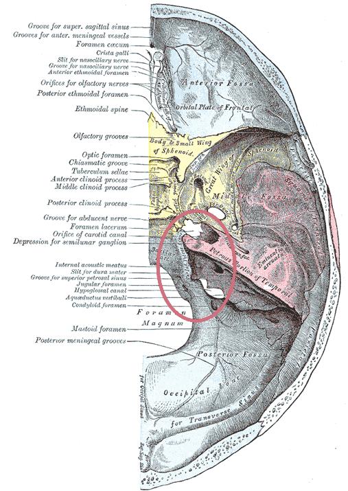
The sutures in the cranial vault are well-known, but the sutures/fissures of the skull base are much less discussed, although they can be classic origins for certain lesions.  One such example is the petro-occipital suture (highlighted below in red; the contralateral suture unmarked), which, not suprisingly, lies between the occipital bone and the petrous aspect of the temporal bone.


The suture links the jugular foramen posteriorly and the foramen lacerum anteriorly.


So why is this suture important?  It is a classic location where chondrosarcomas of the skull base can arise (6% of skull-based lesions).  The painless, locally-invasive tumor arises from remnants of embryonal chondrocytes within the petro-occipital fissure and typically expands upward into intracranial structures.
Radiologically, a destructive mass located at this fissure (heterogeneous enhancement on post contrast T1, hyperintense on T2; chondroid calcification on CT) is highly suspicious of this diagnosis.  The T1 w/ contrast image below (from Reference 2) is an example of a chondrosarcoma arising from the left petro-occipital fissure (the arrowhead demonstrates the petro-occipital fissure on the right).


Incidentally, although the molecular mechanism of eventual petro-occipital suture ossification is similar to that of the cranial vault sutures, it is unclear why it remains relatively unossified in the nonpathological state until late in adulthood.  

A case report of an amyloidoma involving the petro-occipital suture has been described as well (images below). [Reference 3].


It has also been suggested that Maffuci syndrome and Ollier syndrome may be associated with intracranial chondrosarcomas arising from the petro-occipital fissure (Ref. 4).

References:
1. Balboni AL, Estenson TL, et al.  "Assessing Age-Related Ossification of the Petro-Occipital Fissure: Laying the Foundation for Understanding the Clinicopathologies of the Cranial Base." The Anatomical Record Part A 282A:38–48 (2005)
2. Connor SEJ, Leung R, Natas S. "Imaging of the petrous apex: a pictorial review."  The British Journal of Radiology, 81 (2008), 427–435
3. Simoens WA, van den Hauwe L, et al."Amyloidoma of the Skull Base." AJNR Am J Neuroradiol 21:1559–1562, September 2000
4. Tibbs RE, Bowles AP, Raila FA. "Maffuci's Syndrome and Intracranial Chondrosarcoma." Skull Base Surgery Vol 7, no. 1. (1997)