October 30, 2011

The Double Transverse Foramen

The double transverse foramen in the cervical spine is a normal variant that, according to the literature, occurs most often in the C6 vertebra transverse process, with decreasing frequency above and below.  It is claimed that the vertebral artery splits at these accessory foramen (which are always smaller), and rejoins distally, although this is controversial, and it is not entirely clear from the literature whether the accessory foramen usually contains artery, vein, nerve, neither, or some combination. 

Bilateral transverse foramina incidentally noted at C5 on this trauma patient.
Bilateral transverse foramina also incidentally noted at C6 on this trauma patient.  C6 is the most common level to find double transverse foramina.

One source claims that accessory foramina occur in 1.6% of observed cervical spines. The duplication is thought to represent the failure of controlled regression of two intersegmental arteries and a segment of the primitive dorsal aorta.  An extremely rare triple transverse foramen has been reported.


1. Francis CC. "Dimensions of the cervical vertebrae" The Anatomical Record. Anat. Rec. Vol 122: 4 pp.  1097-0185
2. Wysocki J, Bubrowski M, Reymond J, Kwiatkowski J. "Anatomical variants of the cervical vertebrae
and the first thoracic vertebra in man" Folia Morphol. Vol. 62, No. 4, pp. 357–363
3. Murlimanju BV, et al. "Accessory Transverse Foramina in the Cervical Spine: Incidence, Embryological Basis, Morphology and Surgical Importance" Turkish Neurosurgery 2011, Vol: 21, No: 3, 384-387

October 22, 2011

Luckenschadel

Lucke (gap) + schadel (skull)

The luckenschadel (sometimes called "luckenschadel skull," although this is technically redundant) is a calvarial abnormality most often associated with a Chiari II malformation and a meningomyelocele. 

The luckenschadel is an ossification disorder which leads to multiple deep pits in the membranous bones of the newborn skull (favoring the parietal and occipital bones).  It has a classic radiographic "honeycomb" appearance (below), which is diagnostic. 

Alternating regions of cortical fenestration, creating a "honeycomb" appearance, compatible with Luckenschadel.  There is also scalp swelling.  Vaginal delivery of these infants can be problematic since cranial pressure on a fenestrated skull can lead to intracranial hemorrhage.
3D CT reformats of luckenschadel

The grooves in the skull do not necessary traverse the entire bone, and when limited to the inner table, they are called "craniolacunae."  If palpable and involving both the inner and outer table, they are then referred to as "craniofenestrae."  The "waviness" of the skull outline on axial imaging is detectable on obstetric ultrasound (below).


Developmentally, luckenschadel is similar to myelomeningocele, in that there is failure of normal bone formation over the brain.  Some sources claim that it develops in part due to lack of normal cerebral ventricular distention due to an open neural tube defect in the Chiari II malformation.  Over the first 6 months, the fenestrae fill in, and luckenschadel is a diagnosis that can only be made in neonates.


1.   Vigliani, M.  "The luckenschadel skull: a forgotten entity"  Obstetrics and Gynecology: Volume 111(2, Part 2), February 2008, pp 562-565
2.  Coley BD. "Ultrasound diagnosis of luckenschadel (lacunar skull)" Pediatric Radiology, Volume 30:2. pp 82-84.

October 17, 2011

The "Two-eyed Scotty Dog"


The vertebral transverse process contains two additional processes of its own.  The more superior (and larger) of the two is called the mammillary process, which connects posteriorly to the superior articular facet, and is in the same plane as the transverse process.


There is normal variation in the size of this process, but occasionally, a large mammillary process in a sufficiently obliqued radiograph will give the appearance of the "two-eyed scotty dog," with one eye being the usual pedicle and the other eye being the mammillary process.

Oblique view showing the "two-eyed Scotty dog" appearance at L3.  The anterior ring-shaped opacity represent the pedicle (large arrow); the more posterior ring-shaped opacity represents a prominent mammillary process (small arrow).

1.  Patel NP, Kumar R, Kinkhabwala M, Wengrover S. "Radiology of lumbar vertebral pedicles: variants, anomalies, and pathologic conditions"  Radiographics. Vol 7:1 (Jan 1987) pp. 101-137.

October 15, 2011

Torus Torus Torus

50Y M with 60 pack-year history of smoking presents with a painless lump on the hard palate.  Appropriate history taking determines that it has been slowly developing over a long period of time, and the finding can be dismissed as a
torus palatinus, a normal variant.
There are three tori that can potentially occur in the skull: the torus palatinus, the torus maxillaris, and the torus mandibularis.  All three are relatively common (autosomal dominant with variable penetrance) but virtually uneventful bony exostoses.  Case reports of symptomatic tori exist in the literature, but the value of recognizing them on a CT of the skull is essentially to dismiss them as a normal variant.


1.  Gorsky M, Bukai A, Shoha M. "Genetic Influence on the Prevalence of Torus Palatinus" American Journal of Medical Genetics 75:138–140 (1998)
2. Tran KT, Shannon M. "Images in Clincal Medicine: Torus Palatinus"  NEJM 356;17 www.nejm.org april 26, 2007

October 14, 2011

Modic Type 1 change and low back pain

Although controversial, the Modic classification of vertebral endplate changes remains a handy short cut to describe degenerative signal abnormalities.  Originally, two classes were described, the creatively entitled Modic type 1 change and Modic type 2 change.  A rare third Modic type 3 change was added later.  Today's post will focus on the first class: Modic type 1 changes.


Type 1 changes are T1 hypointense and T2 hyperintense.  Like most other areas in the body that exhibit this signal pattern, it is a reflection of increased fluid content in the endplate... signifying bone marrow edema and inflammation.  Type 1 changes exist in a spectrum from Type 1 --> Type 2, indicating an evolution of the degenerative process.  Type 2 can then become active again: Type 2 --> type 1.  If caught at a transition point, the changes are a mixed type 1/2.

T1 sagittal image of the lumbar spine demonstrates a band of decreased signal at the superior  L3 endplate and inferior L2 endplate... the decreased signal is an indication of edema.

T2 sagittal image of the lumbar spine in the same patient demonstrates the same band of endplate signal change, but with increased signal compared to the  T1-weighted images.  The brighter T2 signal is consistent with fluid/edema.

Histologic examination of the endplates in Modic type 1 change show fissuring of the endplates and formation of fibrovascular granulation tissue.  Current opinion is that this change is a result of disc degeneration rather than a factor contributing to disc damage.

The diagnostic dilemma concerning Modic Type 1 changes is that an infectious process (i.e. a discitis) has a similar pattern in the vertebral bodies with inflammation and edema at both endplates adjacent to an infected disc.  The deciding factor is the appearance of the disc itself.  Discitis presents with increased T2 signal intensity, whereas a degenerated, dessicated disc presents with normal or hypointense signal on T2-weighted sequences.  Also, in an infection, other signs are likely to be present, such as erosion of the endplates, a paravertebral soft tissue mass, and appropriate lab tests (CRP (100% sensitive) and ESR).
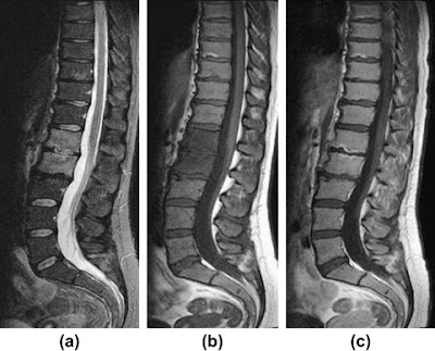
28Y F with  LBP and fever.  Sagittal T2 FS, Sagittal T1 and Sagittal T1+C show L1-L2 discitis and vertebral osteomyelitis.  Note the similar T1 & T2 pattern with Modic type 1 changes.  The difference lies in the disc changes, endplate erosions, and appropriate patient history.

Current opinion also believes that Modic type 1 changes represent a more active form of degenerative change and increased inflammatory cytokines have been recovered from Type 1 vertebral bone.  This Modic type is believed by some to be most closely related to lower back pain, but this remains controversial and studies have been limited by small sample sizes.


1. Rahme R, Moussa R. "The Modic Vertebral Endplate and Marrow Changes: Pathologic significance and Relation to Low Back Pain and Segmental Instability of the Lumbar Spine." AJNR 29 (May 2008)  pp. 838-842.
2. Dunbar JAT, Sandoe AS, et al. "The MRI appearances of early vertebral osteomyelitis and discitis," Clinical Radiology, Volume 65, Issue 12, December 2010, Pages 974-981,

October 13, 2011

Eagle syndrome

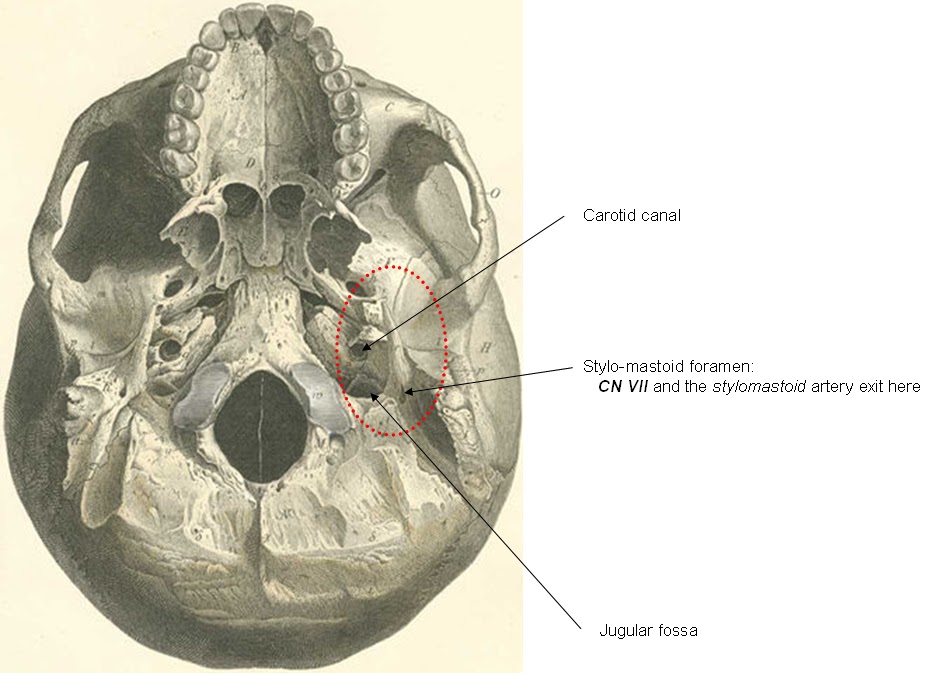
The slender styloid process (Gk. stylus = pillar, instrument for writing (incidentally the root of the word "style")) is seldom seen on most cadaveric skulls since it frequently snaps during dissection.  Technically part of the temporal bone (a.k.a. the "temporal stylus"), the styloid is a point of attachment for multiple small muscles... basically any muscle that originates with the prefix "stylo-" (stylohyoid m., styloglossus m., stylopharyngeal m.).  Just posterior to the styloid process is the stylomastoid foramen, which transmits the exiting CN VII and a small stylomastoid artery.

The skull base ala CT axial views.  The temporal styloid process is circled in red.

Fractures of the styloid have been reported as case reports, but the main contribution of the styloid process to pathology is through Eagle's syndrome in which an elongated styloid process, or a calcified stylohyoid ligament causes chronic neck pain or globus sensation.  The styloid normally measures somewhere around 2-3 cm and in the proper setting, an elongated styloid may contribute to localized pain.  Eagle syndrome can occur unilaterally or bilaterally and cause a variety of nonspecific symptoms including dysphagia, headache, pain on rotation of the neck, pain on extension of the tongue, or dysphonia.  Below is a lateral radiograph of Eagle's syndrome demonstrating a heavily ossified stylohyoid ligament, and beneath that, axial CT images of the same patient.

Heavily calcified structure extending from the skull base to the hyoid is compatible with a stylohyoid ligament calcification (Eagle Syndrome)


Axial CT images of the patient above demonstrate an ossified left stylohyoid ligament.  Compression and entrapment of adjacent structures is not too difficult to imagine with a ossified ligament this flagrant.


One source believes the ultimate source of pathology due to the elongation is entrapment of the glossopharyngeal (CN IX) nerve.


1. Prasad KC, Kamath MP, et al.  "Elongated Styloid Process (Eagle's Syndrome): A clinical study" Journal of Oral and Maxillofacial surgery. 60:2 (Feb 2002) pp. 171-175.
2. Murtagh RD, Caracciolo JT, Fernandez G. "CT Findings associated with Eagle's Syndrome" AJNR Am J Neuroradiol 22:1401–1402, August 2001
3. Slavin KV. "Eagle syndrome: entrapment of the glossopharyngeal nerve? Case report and review of the literature J Neurosurg 97:216–218, 2002

October 5, 2011

The "Human Tail"

Only thirty-three human tails have been reported in the literature, and there is some question how many (if not all) of these represent "pseudotails."  The difference?  A "true" human tail would consist of adipose tissue, skeletal muscle, blood vessels and nerves (no human tail has ever been described containing vertebral bone).  A "pseudotail" is merely a tissue collection (usually a lipoma, teratoma, or myelomeningocele) pouching out at the coccyx with the appearance of a tail (as in the case below).


Human "tails" have been noted to develop in the fetus and then regress.  Case reports note that if the tail persists in the newborn, the finding is very frequently associated with other spinal variations or abnormalities.  Although a tail is certainly not very difficult to detect on a newborn, the history should prompt a careful search of the spine for other spinal abnormalities.

1. Dubrow TJ, Wakym PA, Lesavoy MA. "Detailing the Human Tail" Annals of Plastic Surgery. 20:4 (1988)
2. Alashari M, Torakawa J. "True Tail in a Newborn"  Pediatric Dermatology 12:3 263-266 (1995)